Photomicrograph of honeybee anatomy (please provide details relevant to the part of the anatomy selected)
| Entrant ID: | 1039247 | Entrant Name: | Martin Bullock |
| Entry: | 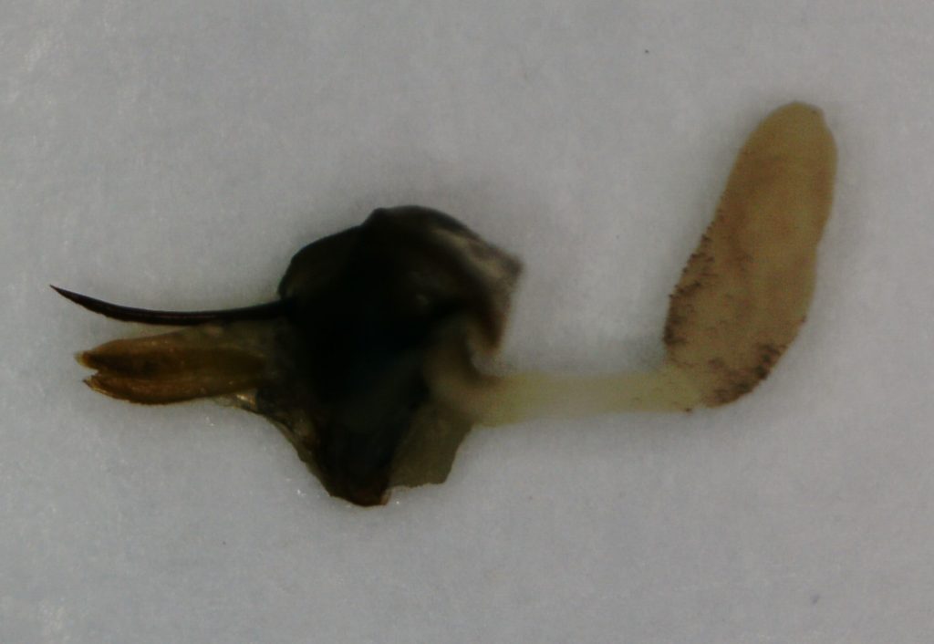 |
||
| Result: | – | ||
| Entrant ID: | 1044039 | Entrant Name: | John MacDougall |
| Description: | Lower front leg of a drone showing part of the basitarsus, 3 tarsomeres, pre-tarsus and foot. The arolium (adhesive pad) and ungues (claws) of the foot are clearly visible. Microscope: Apex Explorer Plus, x40. Camera: Motorola G3 smart phone, hand held. |
||
| Entry: | 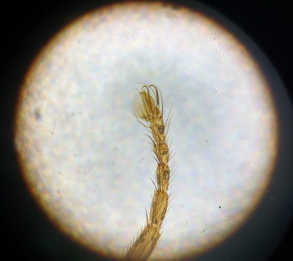 |
||
| Result: |  |
||
| Entrant ID: | 1042567 | Entrant Name: | Gill Brewer |
| Description: | Dissection of queen bee abdomen. X40 When two colonies were united, the spare queen was preserved in isopropanol, for dissection later. For the dissection, the queen was set into beeswax, dorsal side up. A low power microscope, a scalpel, scissors, and needle were used for the dissection. The abdomen was opened by removing the exoskeleton. The massive ovaries, full of hundreds of ovarioles or egg tubes, can be seen on the left. Then the digestive system was lifted out; the rectum with long rectal pads can be seen on the right. Only then, like a pearl, was the spherical spermatheca seen. When a queen bee mates at the beginning of her life, she stores around five million sperm, from the dozen or so drones she mates with, in her spermatheca. This is enough to fertilise all the eggs she lays in her lifetime of up to three or four years. |
||
| Entry: | 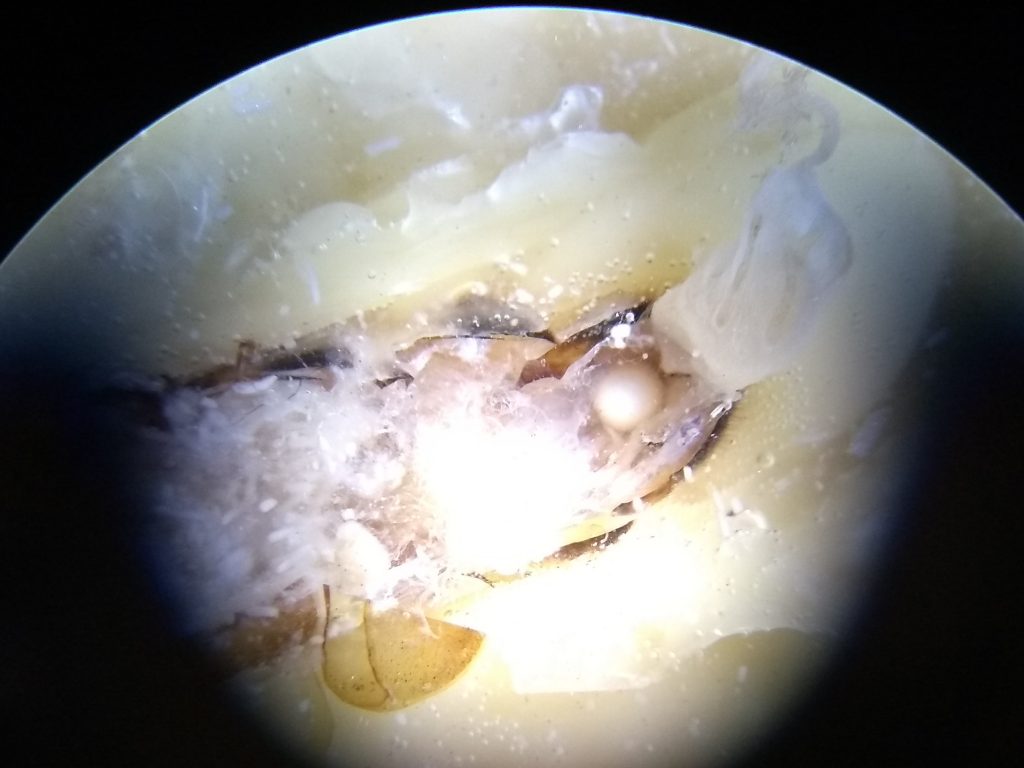 |
||
| Result: |  |
||